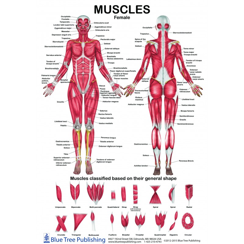
Back Muscles Chart / Greatbigcanvas Labeled Anatomy Chart Of Male Lower Back Muscles On White Background High Definition Acrylic W Charts Posters : Each listed muscle also includes the spinal nerve root level that contributes to the muscles innervation.
Back Muscles Chart / Greatbigcanvas Labeled Anatomy Chart Of Male Lower Back Muscles On White Background High Definition Acrylic W Charts Posters : Each listed muscle also includes the spinal nerve root level that contributes to the muscles innervation.. For more anatomy content please follow us and visit our website: The superior part of the appendicular skeleton that includes clavicle, scapula, and humerus, is attached to the axial skeleton that consists of skull. The extensor muscles are attached to back of the spine and enable standing and lifting objects. If back pain has left you inactive for a long time, a rehabilitation program can help you strengthen your muscles and get back to your daily activities. The most common causes of lower back pain are strain and problems with back structures.
The back anatomy includes the latissimus dorsi, trapezius, erector spinae, rhomboid, and the teres major. To spare you from hours of memorization, we've made some pretty cool video tutorials, articles and quizzes about muscles of the back. If you're going heavy (sets of fewer than about 6 reps), do deadlifts first so you're fresh. A physical therapist can guide you through. A large muscle group in the shoulder, neck and upper back that pulls the head and shoulders backward.

Muscles found in the deep group include the spinotransversales, erector spinae (composed of the iliocostalis, longissimus, and spinalis), the transversospinales, and the segmental muscles.
Related posts of back muscles chart human anatomy muscles leg. Keep your torso upright and a slight arch in your back as you fully extend your arms at the top. Face tmj tongue soft palate ocular middle ear. Pectoral superficial back & scapular arm anterior forearm Three types of back muscles that help the spine function are extensors, flexors and obliques. This helps concentrate more stress on the back muscles. Superficial back muscles, intermediate back muscles and intrinsic back muscles.the intrinsic muscles are named as such because their embryological development begins in the back, oppose to the superficial and intermediate back muscles which develop elsewhere and are therefore classed as extrinsic muscles. Strain commonly occurs with incorrect lifting of heavy. This increases blood flow to the muscle normalizing it and bringing it back to a healthy state. Your clients will thank you for it! If back pain has left you inactive for a long time, a rehabilitation program can help you strengthen your muscles and get back to your daily activities. Shoulders the shoulder muscles are composed of the front, side and rear deltoids. Human anatomy female lower back 12 photos of the human anatomy female lower back human anatomy female lower back, human muscles, human anatomy female lower back.
Head neck upper limb intrinsic back thorax abdomen pelvis & perineum lower limb visceral. These structures work together to support the body, enable a range of movements, and send messages from the brain to. If back pain has left you inactive for a long time, a rehabilitation program can help you strengthen your muscles and get back to your daily activities. Dimitrios mytilinaios md, phd last reviewed: Superficial muscles of the back are located directly deep towards the skin along with superficial fascia.they are occasionally called the appendicular group as these muscles are mainly associated with activities of the appendicular skeleton.

The back anatomy includes the latissimus dorsi, trapezius, erector spinae, rhomboid, and the teres major.
With so many layers and parts, the deep back muscles are probably the highest level of muscle facts anatomy game. This helps concentrate more stress on the back muscles. Human anatomy female lower back 12 photos of the human anatomy female lower back human anatomy female lower back, human muscles, human anatomy female lower back. A physical therapist can guide you through. On this page, you'll learn about each of these muscles, their locations and functional anatomy. Strain commonly occurs with incorrect lifting of heavy. Using proper form and technique on the overhead barbell press targets the deltoids very well for improved strength and size. This procedure is one of the most powerful yet simple ways to treat muscle pain and discomfort. From the chart, you can see the ulnar nerve innervates the flexor carpi ulnaris, abductor digiti minimi, opponens digiti minimi, flexor digiti minimi, lumbricals (3 and 4), interossei, and adductor pollicis muscles. Again, nerve damage associated with these symptoms can be permanent if not treated immediately. October 28, 2020 reading time: Function of the back muscles there are several individual muscles within the back anatomy, and it's important to take a quick look at all of Superficial back muscles, intermediate back muscles and intrinsic back muscles.the intrinsic muscles are named as such because their embryological development begins in the back, oppose to the superficial and intermediate back muscles which develop elsewhere and are therefore classed as extrinsic muscles.
Muscle charts select a region: This includes foot drop, a condition where the muscles of the leg and foot are too weak to raise the foot up as the individual attempts to walk. Human anatomy female lower back 12 photos of the human anatomy female lower back human anatomy female lower back, human muscles, human anatomy female lower back. The top three back training mistakes to avoid [. These structures work together to support the body, enable a range of movements, and send messages from the brain to.

Anatomynote.com found anatomy of back muscles diagram from plenty of anatomical pictures on the internet.
Head neck upper limb intrinsic back thorax abdomen pelvis & perineum lower limb visceral. Human anatomy female lower back 12 photos of the human anatomy female lower back human anatomy female lower back, human muscles, human anatomy female lower back. The seated cable row focuses on both the upper and middle back muscles and is an excellent mass builder. Strain commonly occurs with incorrect lifting of heavy. On this page, you'll learn about each of these muscles, their locations and functional anatomy. The superior part of the appendicular skeleton that includes clavicle, scapula, and humerus, is attached to the axial skeleton that consists of skull. The two trapezius muscles extend from the backbone and base of the skull, across the back and shoulders to join the scapula and the clavicle. If back pain has left you inactive for a long time, a rehabilitation program can help you strengthen your muscles and get back to your daily activities. We are pleased to provide you with the picture named anatomy of back muscles diagram.we hope this picture anatomy of back muscles diagram can help you study and research. This procedure is one of the most powerful yet simple ways to treat muscle pain and discomfort. Artery) p.134 accessory nerve p. These structures work together to support the body, enable a range of movements, and send messages from the brain to. Dimitrios mytilinaios md, phd last reviewed:
Komentar
Posting Komentar